When an operator applies an electric shock of the magnitude of that from a dc defibrillator to the patient’s chest during the T wave of the ECG, there is a strong risk of producing ventricular fibrillation in the patient. Because the most frequent use of defibrillation is to terminate ventricular fibrillation, this problem does not occur; there is no T wave. If, on the other hand, the patient suffers from an atrial arrhythmia, such as atrial tachycardia, fibrillation, or flutter, which in turn causes the ventricles to contract at an elevated rate, dc defibrillation can be used to help the patient revert to a normal sinus rhythm. In such a case, it is indeed possible accidentally to apply the defibrillator output during a T wave (ventricular repolarization) and cause ventricular fibrillation. To avoid this problem, special defibrillators are constructed that have synchronizing circuitry so that the output occurs immediately following an R wave, well before the T wave occurs.
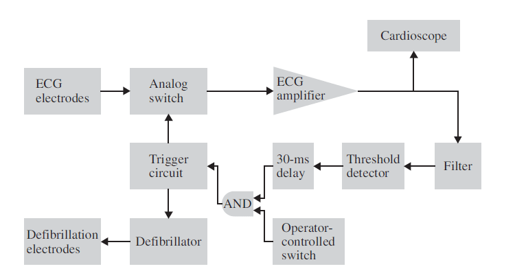
The figure below shows a block diagram of such a defibrillator, which is known as a cardioverter. Basically the device is a combination of the cardiac monitor and the defibrillator. Electrocardiography electrodes are placed on the patient in the location that provides the highest R wave with respect to the T wave. The signal from these electrodes passes through a switch that is normally closed connecting the electrodes to an appropriate amplifier. The output of the amplifier is displayed on a cardioscope so that the operator can observe the patient’s ECG to see, among other things, whether the cardioversion was successful or in extreme cases, whether it produced more serious arrhythmias.

The output from the amplifier is also filtered and passed through a threshold detector that detects the R wave. This activates a delay circuit that delays the signal by 30 msec and then activates a trigger circuit that opens the switch connecting the ECG electrodes to the amplifier to protect the amplifier from the ensuing defibrillation pulse. At the same time, it closes a switch that discharges the defibrillator capacitor through the defibrillator electrodes only once after the operator activates the defibrillator switch. Thus when the operator closes the defibrillator switch, it is discharged immediately after the next QRS complex. After the discharge of the defibrillator, the switch connecting the ECG electrodes to the amplifier is again closed, so that the operator can observe the cardiac rhythm on the cardioscope to determine the effectiveness of the therapy.
You can also read: Principle Parts and Types of ECG Recorders

Leave a Reply
You must be logged in to post a comment.