Magnetic resonance imaging (MRI) is a medical imaging modality which employs magnetic fields and radio waves/radiofrequency (RF) energy to produces images of the body. This imaging technique is based on nuclear magnetic resonance (NMR), which is a quantum mechanical phenomenon exhibited by atoms having either an odd number of protons or neutrons. Such atoms have nonzero nuclear magnetic moment, μ, and will precess (or rotate) about an external magnetic field (B0) with a frequency of ω0 = ϒB0 where ϒ is the gyromagnetic ratio which for 1H is 42.57 MHz/T. While several isotopes such as 1H, 23Na, 31P and 2H exhibit NMR phenomena, most of the MRI scanners use 1H. This is due to the fact compared to other isotopes 1H has a high inherent sensitivity and abundance in tissue. When placed in a B0 field 1H nuclei aligns their spins either parallel or antiparallel to B0 with a slight excess in the lower energy parallel state.
Key Components of a Modern MRI Scanner
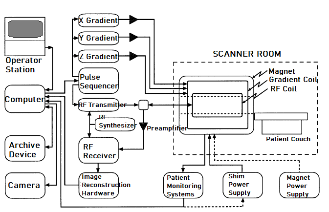
The key components of a modern MRI system include a magnet, a pulse sequencer, gradient & shim coils and an RF transmitter/receiver – the function of which are controlled by a host computer. These components are illustrated in the figure below:

In reference to the image above, to obtain the magnetic field (B0) most MRI scanners supplied by vendors use superconducting magnets, however, some special-purpose (and often lower-cost) scanners may use resistive magnets. Superconducting magnets typically have field strengths between 0.5 and 4.0 T, while resistive magnets normally have field strengths < 0.3 T. The enhanced signal-to-noise ratio obtained with superconducting designs is offset by the requirement for periodic cryogen replacement. Most modern magnets, however, minimize this cost by including a cryogen reliquefier.
Two sets of auxiliary gradient coils are positioned within the main magnet to provide spatially varying fields, and to allow shimming of the B0-field. Gradient coil hardware in modern MRI scanners can generate maximum gradients of up to 40 mT/m with rise times of 120 μs and allow fields of view of 4 to 48 cm. Shim coils enhance the homogeneity of the B0 field by decreasing δB0(r) [i.e. reversible field inhomogeneity due to imperfect magnet] to a few parts per million in order to minimize effects of relaxation time (T2) that characterizes spin-spin relaxation process and also lower spatial distortions.
Modern MRI scanners include a digital RF subsystem which excites the spins and then records the emitted signals via one or more RF coils within the magnet. An RF synthesizer is coupled to both the RF transmitter and receiver, so that synchronous detection is possible. The RF system is connected either to separate transmit and receive coil(s) or to a combined transmit/receive coil(s).
To obtain an MRI image, the host computer interacts with the operator who defines the imaging parameters such as the angle in the y-z plane (α), echo time (TE) and repetition-time (TR), slice location and field of view. The parameters are then translated into instructions which are executed on a synchronous state machine referred to as a pulse sequencer. This device provides a real-time control of the gradient and RF waveforms as well as other control functions, such as the un-blanking the RF receiver and enabling the analog-to-digital converter (ADC) during an echo. The data collected is collected and demodulated by the receiver, and then images are reconstructed using specialized hardware built typically around a fast-array processor. The images are sent to the host computer for operator station display, archival or filming.
Find out more about: Fingertip Pulse Oximeter Blood Oxygen Saturation Monitor
It is important to also note that, most MRI scanners include devices for monitoring heart and respiration rate, and allow these signals to trigger or gate image acquisition.
More research and innovations in MRI imaging systems may make it possible to incorporate higher B0 field and gradients, and also achieve faster data processing or reconstruction hardware.
Related articles:
- Magnetic Resonance Imaging (MRI)
- What is Functional MRI (fMRI)?
- Radionuclide Imaging Techniques
- Positron Emission Tomography (PET)
- Ultrasound Scanning Techniques
- X-ray Imaging
- What is CAT scan?

Leave a Reply
You must be logged in to post a comment.